


Chapter 6 Microscopy Applications 47
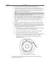
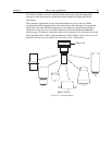
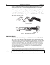
Adjusting the Parfocality of the Camera
On a C-mount system, the camera should be very close to parfocal, although some
C-mounts will be adjustable using set screws on the microscope to secure the adapter
slightly higher or lower in position.
To adjust the parfocality on an F-mount system, begin collecting images with a short
exposure time and focus the light on the camera by rotating the ring on the Relay Lens
without touching the main focusing knobs on the microscope.
While adjusting parfocality, you will need to acquire images rapidly to minimize the
delay between the time a setting is changed and the time when the effect of the change
can be observed. The specifics of how to proceed will vary according to the application
software.
In WinView, select Acquisition, Focus. Begin with an exposure time 0.1 sec. Then
use RUN to begin data acquisition and STOP to end it when you are finished focusing.
See your WinView manual for additional information.
Many Princeton Instruments cameras, both C-mount and F-mount, also make provision
for extending the focus range by providing a focus adjustment on the camera lens mount.
If necessary, this focus can be changed to bring the image into range of the relay lens or
other microscope focus adjustment as described on page 41.
Imaging Hints
Determine the gray levels of the image by placing the cursor within the image and
monitoring the values shown. For optimal image quality of a 12-bit image, the highest
value in the field should be over 3000 counts but less than 4095 (which is saturating).
You may increase the number of counts by increasing your exposure or by increasing the
amount of light illuminating the specimen.
Fluorescence
Once you have acquired a suitable image in transmitted light mode, you may switch to
fluorescence mode.
In fluorescence mode you generally want to minimize the bleaching of your sample,
usually achieved by placing several neutral density filters in the excitation pathway to
minimize the illumination intensity. There will always be a trade-off here; when you
maximize signal quality by increasing the illumination intensity, you need to consider
whether your preparation can tolerate these conditions. In general, it is better to expose
longer with a lower intensity than to expose for a shorter time with a higher intensity;
nevertheless, your experimental conditions will dictate which path you take.
In fluorescence measurements you may not wish to maximize the gray levels in the
image, since this may cause bleaching of the dye or photodamage to the cell. Maintain
the minimum exposure required to get a sufficiently high quality image.
If the scaling on the image does not appear good to the eye, you may use additional
scaling features available in the software. See your software manual for information on
how to properly use the contrast enhancing features of the program.